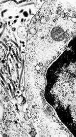
The electron micrograph on the left is of Ebola Zaire in a lymph node of
an African
green monkey. It was done by Dr. Tom Geisbert.
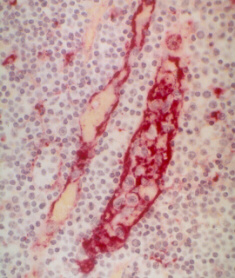
The image on the right is of a lymph node of an infected African Green
Monkey.
The location of the Ebola Zaire antigen is indicated by the red
stain.
The large ovoid structure at the center of the picture is a high
endothelial venule (HEV) that is infected with ebola. The viral
replication in the fibroblastic cells that control the HEV's
structure
has almost totally destroyed the HEV. Please see Dr. Art Anderson's
web page for a more detailed explanation for this interpretation.
This
image was prepared by Art Anderson from one of Keith Steele's slides
of ebola antigen IHC.
Dr. Art Anderson took this photomicrograph of the lip of an African
green monkey. Ebola virus is penetrating between the epithelial cells
of the lip overlying a lymphoid aggregate in the submucosa. This
immunohistochemistry preparation was done by Keith Steele.
Dr. Art Anderson graciously supplied these pictures and their explanations.
For a more detailed analysis of this characteristic of Ebola pathogenesis,
please see:
Dr.
Art Anderson's Explanation of Lymphocyte Homing